Bringing Interferometric Imaging into the X-Ray Regime
[ad_1]
• Physics 16, 66
The experimental realization of a just lately proposed method factors to new prospects for imaging molecules utilizing x rays.
Stacy Huang
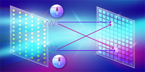
Hanbury Brown and Twiss (HBT) interferometry [1] is a flexible method broadly utilized in numerous fields of physics, similar to astronomy, quantum optics, and particle physics. By measuring the correlation of photon arrival occasions on two detectors as a perform of the photons’ spatial separation, HBT interferometry permits the dedication of the dimensions and spatial distribution of a light-weight supply. Lately, a novel x-ray imaging method based mostly on the HBT technique was proposed to picture the spatial association of heavy parts in a crystal or molecule by inducing these parts to fluoresce at x-ray wavelengths [2]. Now Fabian Trost of the German Electron Synchrotron (DESY) and colleagues—together with a few of those that first proposed the scheme—have applied this method, efficiently demonstrating that the temporal correlation of fluorescence photons on a detector can be utilized to picture the construction of emitters on a copper movie [3]. This achievement marks a big milestone towards extending HBT interferometry into high-resolution x-ray imaging, with the potential to picture the construction and dynamics of remoted biomolecules with out the necessity for crystallization [4].
The HBT impact is a two-photon interference phenomenon that happens when two indistinguishable photons emitted from totally different factors inside a supply attain two totally different detectors. The indistinguishability situation is happy when the time interval between the 2 photons is inside the coherence time of the sunshine supply. The interference impact, whether or not constructive or harmful, will be quantified utilizing the second-order correlation perform, or g(2), which describes the likelihood of detecting two photons concurrently as a perform of their spatial separation. If the arrival time is longer than the coherence time, this can result in decreased distinction within the interference fringes in g(2).
Extending the HBT method into the x-ray regime has been a problem because of the low likelihood of x-ray excitation and the quick coherence time of x-ray fluorescence, particularly for heavy parts. Right here the coherence time is given by the lifetime of the fluorescence states. For instance, the lifetime of the digital state of a copper atom with a emptiness within the Ok shell is lower than 1 fs. The emission from two copper atoms will solely be coherent or indistinguishable if the atoms are excited inside that interval. The event of x-ray free-electron lasers (XFELs) has helped on this regard by making doable the technology of high-intensity x-ray pulses with femtosecond and even shorter durations [5]. These pulses can excite the Ok-shell electrons of heavy parts with a excessive likelihood and enhance the prospect of manufacturing indistinguishable pairs of photons by way of x-ray fluorescence.
Trost and colleagues utilized such pulses to measure the depth correlation of fluorescence photons emitted from a copper movie. Conducting their experiment on the European XFEL facility in Germany, the researchers used a part grating to diffract the incoming x-ray pulse and focus it onto two spots on a micron-sized copper foil. The incident x-ray photons had an vitality of 9 keV, which was adequate to ionize the Ok-shell electron of the illuminated copper atoms on the movie. This ionization course of created a short-lived excited state that primarily decayed by way of fluorescence emission, which the researchers measured utilizing a detector specifically developed on the XFEL facility. With a million pixels, every able to single-photon detection, this detector can measure 1012 correlations between pixel pairs.
The crew wanted to deal with a number of challenges as a way to implement x-ray imaging utilizing this method. One problem was that the detector used within the experiment doesn’t resolve the vitality of the photons, which means it can not exclude contributions from different sources of radiation in addition to the specified emission. This nonselectivity would result in a low signal-to-noise ratio. To handle this situation, Trost and colleagues used a nickel filter to dam elastically scattered radiation and copper radiation.
One other problem was that the length of the x-ray pulse was 10 occasions longer than the coherence lifetime of the emission, which reduces the distinction of the interference fringes. To enhance the sign high quality, the researchers recorded 58 million 2D fluorescence photos in about 5 hours, enabled by the detector’s excessive readout charge and by the excessive repetition charge of the XFEL pulses. To make sure that the pattern wasn’t broken from the pulses, the copper foil was rotated such that every pulse illuminated a brand new space. Though the mixed fluorescence picture was isotropic and contained no structural data, the constructed g(2) revealed interference fringes reaching the third-order peaks, which is an enchancment in comparison with earlier research that solely measured the zero-order peak in g(2) [6, 7]. By utilizing an iterative algorithm, the researchers efficiently reconstructed the dimensions (300 nm) and separation (860 nm) of the 2 excited spots on the copper movie.
Given substantial enchancment in spatial decision, the method might finally allow single-particle imaging of biomolecules and catalysts on the atomic scale, and, with adequate time decision, characterization of their response dynamics. Continued advances in XFEL know-how might additionally imply that the photon-hungry means of fluorescence excitation will be achieved with subfemtosecond pulses. For instance, utilizing intense, subfemtosecond x-ray pulses, it’s doable to tailor the temporal fluorescence profile to be shorter than the lifetime of the fluorescing states [8]. To take advantage of the ability of HBT interferometry for chemical imaging with elemental specificity, it’s extremely fascinating to develop multicolor imaging utilizing a number of detectors or detectors with energy-discrimination capabilities. Additional improvement on this respect has the potential to revolutionize the characterization of essential catalytic features and of the related structural modifications of their native environments—similar to metal-bearing clusters in metalloproteins [9]. Such characterization would offer unprecedented perception into these catalysts’ construction and exercise, which is vital for the event of renewable vitality sources.
References
- R. Hanbury Brown and R. Q. Twiss, “Correlation between photons in two coherent beams of sunshine,” Nature 177, 27 (1956).
- A. Classen et al., “Incoherent diffractive imaging by way of depth correlations of arduous x rays,” Phys. Rev. Lett. 119, 053401 (2017).
- F. Trost et al., “Imaging by way of correlation of x-ray fluorescence photons,” Phys. Rev. Lett. 130, 173201 (2023).
- R. Neutze et al., “Potential for biomolecular imaging with femtosecond X-ray pulses,” Nature 406, 752 (2000).
- J. Duris et al., “Tunable remoted attosecond x-ray pulses with gigawatt peak energy from a free-electron laser,” Nat. Photonics 14, 30 (2019).
- I. Inoue et al., “Willpower of x-ray pulse length by way of depth correlation measurements of x-ray fluorescence,” J. Synchrotron Radiat. 26, 2050 (2019).
- N. Nakamura et al., “Focus characterization of an x-ray free-electron laser by depth correlation measurement of x-ray fluorescence,” J. Synchrotron Radiat. 27, 1366 (2020).
- P. J. Ho et al., “Fluorescence depth correlation imaging with excessive spatial decision and elemental distinction utilizing intense x-ray pulses,” Struct. Dyn. 8, 044101 (2021).
- J. Yano and V. Yachandra, “Mn4CA cluster in photosynthesis: The place and the way water is oxidized to dioxygen,” Chem. Rev. 114, 4175 (2014).
Concerning the Writer
Topic Areas
[ad_2]


